3D/4D Ultrasound Technology
3D/4D vaginal ultrasonography in fertility care: seeing is beyond believing!
By Patsama Vichinsartvichai, MD., MClinEmbryol. Medical Director. Inspire IVF
At Inspire IVF, we’re well aware that every step along your fertility journey is crucial and will be a memory of your lifetime.
The latest in 3D/4D vaginal scan Ultrasound Technology is provided to you, by Inspire IVF, as a part of your care.
This technology is lead by Thailand’s national expert in Gynecologic Sonography, our Medical Director, Dr Pat. So you can be sure that no single step in your diagnosis is missed.
In contemporary infertility care, vaginal sonography is commonly used in the diagnosis, monitoring, and treatment in infertility (Figure 1).
 Figure 1 Fertility treatment cycle that vaginal sonography involves
In recent years, the technology of ultrasound has advanced tremendously. Some brands release new models every year like iPhone!
With the integration of piezoelectric materials into 2D ultrasound technology, the 3D/4D vaginal scan that we use now has become the most powerful tool in fertility treatment, since it can be applied effectively to every step of care1.
Cycle monitoring
Cycle monitoring by measurement of the size of follicle can achieve much quicker and much more accurate results with 3D/4D vaginal scan. The software will pick up the follicle and measurement. Another confirmation of size and location of follicles is done by the doctor to make sure there’s nothing missed.(Figure 2)1, 2.
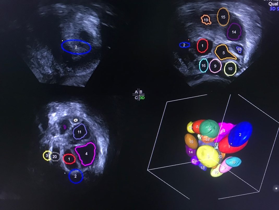
Figure 2 An ovary in volume analysis mode picturing individual growing follicle measurement.
First trimester fetal measurement and nuchal translucency analysis
After a pregnancy test is confirmed positive, our doctor will make an appointment with you to scan the health of the fetus every two weeks to confirm the fetal heart activity and the growth of the fetus.
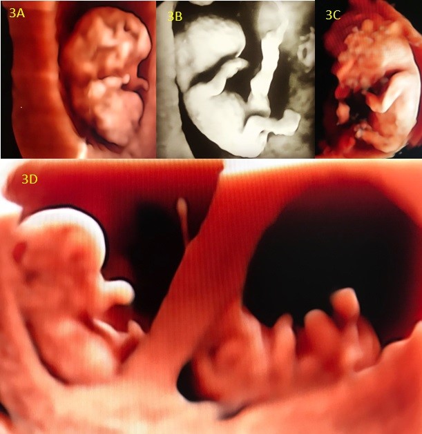
3D/4D vaginal scan makes the picture of your fetus more realistic and heartfelt. The nuchal translucency scan, which relates to chromosomal aneuploidy such as Downs Syndrome, can be more accurately measured with 3D/4D vaginal scan, rather than regular 2D ultrasound which depends on the position of the fetus.

Figure 3 3D/4D vaginal scan of first trimester fetuses with HDlive rendering mode A. an 8 weeks fetus B. 10 weeks fetus C. 12 weeks fetus. He turns his back toward the probe so the image of facial profile can’t be seen. D. a set of twins at 8 weeks after transferred 2 blastocysts.
Diagnosis
The causes of female infertility can be anatomical, ovulatory disorder, chemical inflammation, and immunologic disorders (Figure 2)2.

Figure 4 Causes of female infertility
In recent publications by Dr Patsama, he found that endometriosis causes around 20-30% of infertility in Thai women and most of them have severe stage of disease3, 4.
The 3D/4D vaginal scan helps the doctor to evaluate the depth, length and width of deep endometriosis involving the rectum, before surgery, to help the patient have complete surgery for the best outcomes (Figure 3A and 3B).
Ultrasonography has benefits over MRI since it can give real-time imaging of organ relationships and adhesion. With current technology, the depth of lesion can also determined and the treatment can be planned properly.
Figure 5A and 5B: A set of 3D/4D vaginal sonography of deep endometriosis involving the uterus and rectum 3A. A multiplanar mode of 3D vaginal sonography demonstrates the length, width and depth of deep endometriosis . 3B. Rendered image of the deep endometriosis lesion.
In patients with ovulatory disorder such as PCOS, 3D/4D vaginal scan helps in counting the antral follicles (AFC) and makes it more accurate than a manual count (Figure 4). The ovarian volume can also be measured, which helps with prediction of disease and androgen status5.

Figure 6 A volume calculation scan of an ovary of a patient with PCOS during stimulating cycle demonstrating over 50 growing follicles in one ovary. This might take over 30 minutes to measure the follicle with 2D ultrasound.
Congenital uterine anomaly and intracavitary lesion can lead to infertility as well.
Traditionally, office hysteroscopy is performed to evaluate the uterine cavity, even if it’s not painful and no cervical dilatation is needed.
With 3D/4D vaginal scan, the uterine cavity can be evaluated in a minute or less without pain just like 2D vaginal sonography6.
With our experts performing the scan, the performance of 3D/4D vaginal scan to diagnose uterine anomalies is non-inferior to MRI. 3D/4D vaginal scan gives you a new sonographic view that only MRI could perform before “coronal view” which we use in diagnosis of the uterine cavity.
We might call this virtual hysteroscopy!


Figure 7 Demonstrates various type of uterine anomaly diagnosed from coronal view of 3D/4D vaginal scan. 5A. normal uterine cavity, 5B. partial septate uterus 5C. Hypoplastic T-shape uterus, 5D. Arcuate uterus 5E. Another hypoplastic uterus.
References
- Huang Q, Zeng Z. A Review on Real-Time 3D Ultrasound Imaging Technology. Biomed Res Int. 2017;2017:6027029.
- Recent advances in medically assisted conception. Report of a WHO Scientific Group. World Health Organ Tech Rep Ser. 1992;820:1-111.
- Vichinsartvichai P, Siriphadung S, Traipak K, Promrungrueng P, Manolertthewan C, Ratchanon S. The influence of women age and successfulness of intrauterine insemination (IUI) cycles. Journal of the Medical Association of Thailand. 2015;98.
- Vichinsartvichai P, Traipak K, Manolertthewan C. Performing IUI simultaneously with hCG administration does not compromise pregnancy rate: A retrospective cohort study. Journal of Reproduction and Infertility. 2018;19.
- Sujata K, Swoyam S. 2D and 3D Trans-vaginal Sonography to Determine Cut-offs for Ovarian Volume and Follicle Number per Ovary for Diagnosis of Polycystic Ovary Syndrome in Indian Women. J Reprod Infertil. 2018;19:146-51.
- Apirakviriya C, Rungruxsirivorn T, Phupong V, Wisawasukmongchol W. Diagnostic accuracy of 3D-transvaginal ultrasound in detecting uterine cavity abnormalities in infertile patients as compared with hysteroscopy. Eur J Obstet Gynecol Reprod Biol. 2016;200:24-8.
- Feng Y, Tamadon A, Hsueh AJW. Imaging the ovary. Reproductive BioMedicine Online. 2018;36:584-93.

